Oncos-Molbiol-Logo

Diagnosis

❐ Cancer Screening

Diagnosing cancer at its earliest stages often provides the best chance for a cure. With this in mind, talk with your doctor about what types of cancer screening may be appropriate for you. For a few cancers, studies show that screening tests can save lives by diagnosing cancer early. For other cancers, screening tests are recommended only for people with increased risk.

A variety of medical organizations and patient-advocacy groups have recommendations and guidelines for cancer screening. Review the various guidelines with your doctor and together you can determine what’s best for you based on your own risk factors for cancer.

The focus of cancer control is to diagnose cancer in the earliest possible curable stage, thus improving the quality of life and prognosis of cancer patients.

WHO summarized the major elements for cancer early diagnosis into three main domains:


Step 1: Awareness of cancer symptoms and accessing care

Step 2: Clinical evaluation, diagnosis, and staging

Step 3: Access to treatment, including pain relief

❐ Cancer Diagnosis

There are several methods of diagnosing cancer. With advances in technologies that understand cancers better, there is a rise of number of diagnostic tools that can help detect cancers. Once suspected, diagnosis is usually made by pathologists and oncopathologists and imaging radiologists.

Some types of cancer, particularly lymphomas, can be hard to classify, even for an expert. Most cancers need a second opinion regarding diagnosis before being sure of the diagnosis or stage and type.

The most common diagnostic methods include:

⯐ Physical Exam

Your doctor may feel areas of your body for lumps that may indicate cancer. During a physical exam, your doctor may look for abnormalities, such as changes in skin color or enlargement of an organ, that may indicate the presence of cancer.

⯐ Laboratory Tests

Laboratory tests, such as urine and blood tests, may help your doctor identify abnormalities that can be caused by cancer. For instance, in people with leukemia, a common blood test called complete blood count may reveal an unusual number or type of white blood cells. Blood tests can be performed to detect the normal blood cells as well as for specific tumor markers. Some tumors release substances called tumor markers, which can be detected in the blood. A blood test for prostate cancer determines the amount of prostate specific antigen (PSA). Higher than normal PSA levels can indicate cancer. Similarly in ovarian cancer a tumor marker CA-125 is released..

❐ Biopsy

This is a test where a small sample of tissue is taken from the suspected cancer with the help of a fine tipped needle (fine needle aspiration – FNA), or with a thicker hollow needle (core biopsy) or by surgical excision. The tissues are then examined under a microscope for the presence of cancer cells. Depending on tumor location, some biopsies can be done on an outpatient basis with only local anesthesia.

❐ Sentinel Node Biopsy

This is a procedure where the closest and most important nodes near the cancer are surgically excised and examined. Since sentinel nodes are the first location that cancer is likely to spread, only these lymph nodes that likely contain cancer cells.

❐ Endoscopy

In this imaging technique a thin, flexible tube with a tiny camera on the end is inserted into the body cavities. This allows the doctors to view the suspicious area. There are many types of scopes, each designed to view particular areas of the body. For example, a colonoscopy looks at the colon and large intestine and a laparoscope is used to look within the abdomen etc.

❐ Bone Marrow Aspiration

These show a picture of the bone marrow that may be affected in leukemia and blood cancers.

❐ Pap Test

Pap test (Pap smear) is a routine test where a sample of cells from a woman’s cervix is examined under the microscope. This helps identify changes in the cells that could indicate cervical cancer or other conditions.

❐ Sputum Analysis and Bronchial Washing Analysis

The cells of the sputum and bronchial secretions are analyzed under the microscope for signs of lung and other respiratory cancers.

❐ Imaging Studies

There are several imaging techniques. These include X rays, CT scans, MRI scans of various parts of the body.

X-rays are the most common imaging techniques and they may be made more specific by using a Barious enema. This is used for detection of stomach and small intestinal growths and cancers.

Mammogram is an X-ray of the breasts used to screen for and/or detect breast lumps and growths.

A CAT scan (computerized axial tomography) uses radiographic beams to create detailed computerized pictures. It is more precise than a standard X-ray.

An Magnetic Resonance Imaging (MRI) uses a powerful magnetic field to create detailed computer images of the body’s soft tissue, large blood vessels and major organs. Both CT scan and MRI can also be used with contrast radio-labelled dyes to obtain a more clear and specific picture of the cancer.

An Ultrasound uses high-frequency sound waves to determine if a suspicious lump is solid or fluid. These sound waves are transmitted into the body and converted into a computerized image.

Bone scan is specifically used to identify and locate new areas of cancer spread to the bone. Normally a Positron imaging test (PET scan) is used. A Gallium scan is another nuclear medicine test in which a special camera takes pictures of tissues of the body after a special radioactive tracer is injected into a vein. The cancerous areas light up under the scanner.

❐ Genetic Analysis

Cytogenetic analysis involves analysis of blood or bone marrow cells for organizations of chromosomes. This shows up any genetic mutations.

❐ Cytological Methods

These methods for diagnosis consist of study of cells shed off into body cavities and study of cell by putting a fine needle introduced under vacuum into the lesion(fine needle aspiration cytology FNAC).

❐ Fine Needle Aspiration Cytology

Fine Needle Aspiration Cytology (FNAC) is a diagnostic procedure where a needle is inserted into your body, and a small amount of tissue is sucked out for examination under a microscope.

It is a quicker and less painful procedure than a Biopsy, but doctors still prefer a biopsy for lymphomas as it gives surer results.

❐ Application

This type of sampling is performed for one of two reasons

1.  A biopsy is performed on a lump or a tissue mass when its nature is in question.

2.  For known tumors, this biopsy is performed to assess the effect of treatment or to obtain tissue for special studies.

When the lump can be felt, the biopsy is usually performed by a Cytopathologisticor a Surgeon. In this case, the procedure is usually short and simple. Otherwise, it may be performed by an interventional radiologistic, a doctor with training in performing such biopsies under X-ray or ultrasoundguidance. In this case, the procedure may require more extensive preparation and take more time to perform.

Also, fine-needle aspiration is the main method used for chorionic villus sampling as well as for many types of body fluid sampling, as shown in Fig 1and Fig 2.

Fig. 1: FNAC method for taking of samples from tissues


Fig. 2: FNAC method for taking of samples from thyroid tissues


❐ Histochemistry and Cytochemistry

These are additional diagnosis tools which helps the pathologist in identifying the chemical composition of cell.Their constituents and their products by special staining methods.

Though immunohistochemical techniques are more useful for tumourdiagnosis, histochemical and cytochemical methods are still employed for this purpose.

❐ Immunohistochemistry

This is an immunological method of recognizing a cell by one or more of its specific components in the cytoplasm,cell membrane or nucleus . These cell components are combine with specific antibodies on the formaline fixed paraffinesection.the complex of antigen and antibody on the slide is made visible for light microscopic identification by either fluorescent dyes or by enzymes, as shown in Fig 3.

The list of immunochemical stains is ever increasing; one important group of such antibody stains is directed against various classes ofintermediate filaments which is useful in classification of poorly-differentiated tumours of epithelial or mesenchymal origin. list of tumours and stains are shown in Table 1.

❐ Electron Microscopy

1.  Ultra structural examination of tumour call offers selective role in diagnostic pathology

2. Cell junctions-their presence and type.

3. Cell surface eg: presence of microvilli.

4. Cell shape and cytoplasmic extinction.

5. Shape of the nucleus and features of nuclear memberane.

6. Nucleoli-size and density.

7. Cytoplasmicorgenelles-their number is generally reduced.

8. Dense bodies in the cytoplasm.

9. Any other secretory products in the cytoplasameg: melanosomesin melanoma and memberane bound granules in endocraintumours.

❐ Tumour Markers

Tumor markers are substances that are produced by cancer or by other cells of the body in response to cancer or certain benign (noncancerous) conditions. Most tumor markers are made by normal cells as well as by cancer cells; however, they are produced at much higher levels in cancerous conditions. These substances can be found in the blood, urine, stool, tumor tissue, or other tissues or bodily fluids of some patients with cancer. Most tumor markers are proteins. However, more recently, patterns of gene expressionand changes to DNA have also begun to be used as tumor markers. Markers of the latter type are assessed in tumor tissue specifically.

Thus far, more than 20 different tumor markers have been characterized and are in clinical use. Some are associated with only one type of cancer, whereas others are associated with two or more cancer types. There is no “universal” tumor marker that can detect any type of cancer.

Tumor markers are used to help detect, diagnose, and manage some types of cancer. Although an elevated level of a tumor marker may suggest the presence of cancer, this alone is not enough to diagnose cancer. Therefore, measurements of tumor markers are usually combined with other tests, such as biopsies, to diagnose cancer.

A doctor takes a sample of tumor tissue or bodily fluid and sends it to a laboratory, where various methods are used to measure the level of the tumor marker. List of tumours markers for different cancers are listed in Table 2.

❐ Flow Cytometry

In biotechnology, flow cytometry is a laser- based, biophysical technology employed in cell counting, cell sorting, biomarkers detection and protein engineering, by suspending cells in a stream of fluid and passing them by an electronic detection apparatus. It allows simultaneous multipara metric analysis of the physical  and chemical characteristics of up to thousands of particles per second.

Flow cytometry is routinely used in the diagnosis of health disorders, especially blood cancers, but has many other applications in basic research, clinical practice and clinical trials. A common variation is to physically sort particles based on their properties, so as to purify populations of interest.

Flow cytometry is a technology that is used to analyses the physical and chemical characteristics of particles in a fluid as it passes through at least one laser. Cell components are fluorescently labeled and then excited by the laser to emit light at varying wavelengths.

The fluorescence can then be measured to determine the amount and type of cells present in a sample. Up to thousands of particles per second can be analysed as they

pass through the liquid stream.Which is shown in Fig 4 and Fig 5.

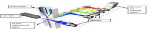
Fig. 4: Flow cytometry


Fig. 5: Graphical representation of different components detected by using flow cytometry


Applications

This laser-based technology is used to perform several procedures including:

Cell counting

Cell sorting

Detection of biomarkers

Protein engineering

Flow cytometry has numerous applications in science, including those relevant to healthcare. The technology has been widely used in the diagnosis of health conditions, particularly  diseases   of   the   blood   such as leukemia, although it is also commonly used in the various different fields of clinical practice as well as in basic research and clinical trials.

Some examples of the fields this technology is used in include molecular biology,immunology, pathology, marine science and plant biology. In medicine, flow cytometry is a vital laboratory process used in transplantation, oncology, hematology, genetics and prenatal diagnosis. In marine biology, the abundance and distribution of photosynthetic plankton can be analysed. Size of particals is 1-50micron size. Flow cytometry can also be used in the field of protein engineering, to help identify cell surface protein variants.

This is a computerized technique by which the detailed characteristics of individual tumour cell are recognized and quantified and the data can be stored for subsequent comparison too.

❐ In Situ Hybridization

This is a molecular technique by which nucleic acid sequences can be localized by specifically labeled nucleic acid probe directly in the intact cell rather than by DNA extraction. It is shown in Fig 6.


Fig. 6: Insitu hybridizations

❐ Molecular Diagnostic Technique:

The group of molecular biologic methods in the tumour diagnostic laboratory are a variety of DNA/RNA-based molecular technique in which the DNA/RNA are extracted from the cell and then analysed.the molecular methods in tumour diagnosis can be applied in hematologic as well as non-hematologic malignancies by

–          Analysis of molecular cytogenetic abnormalities.

–          Mutational analysis.

–          Antigen receptor gene rearrangement and

–          By study of the oncogenic viruses at molecular level.

DNA Microarray Analysis of Tumours Currently, it is possible to perform molecular profiling  of  a  tumour  by  use of  gene                chip technology  which  allows  measurement of level of expression of several thousand genes simultaneously.

The core principle behind microarrays is hybridization between two DNA strands, the property of complementary nucleic acid sequences to specifically pair with each other by forming hydrogen bonds between complementary nucleotide. A high number of complementary base pairs in a nucleotide sequence mean tighter non-covalent bonding between the two strands. After washing off non-specific bonding sequences, only strongly paired strands will remain hybridized. Fluorescently labeled target sequences that

bind to a probe sequence generate a signal that depends on the hybridization conditions (such as temperature), and washing after hybridization. Total strength of the signal, from a spot (feature), depends upon the amount of target sample binding to the probes present on that spot. Microarrays use relative quantitation in which the intensity of a feature is compared to the intensity of the same feature under a different condition, and the identity of the feature is known by its position. It is observed in Fig 7,8.


DNA microarray technology is a promising approach that allows both qualitative and quantitative screening for sequence variations in the genomic DNA of cancer cell. DNA microarray based samples.

x
Start Chat
Need help?
Scan the code
Hello 👋
How can we help you?